Table of Contents
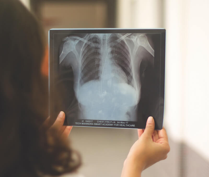
ToggleRadiology is a branch of medicine that uses imaging technology to diagnose pathological conditions and guide in therapeutic procedures. Medical Imaging tools act as a camera to take pictures of internal organs. The way photos quality depends on the types of cameras used, photographers and developing conditions. Similarly, radiological image quality depends on characteristics of imaging equipment, skill of the radiological technician, radiation exposure to the patient, and imaging time.
Five major components of medical imaging process are –
- The object in the patient body
- The imaging equipment
- Radiographer
- Image itself
- The observers or radiologist who will conclude the study
Quality Imaging
This is the fourth component of the Medical Imaging process. Quality Imaging refers to the fidelity with which the anatomic structures being examined are imaged on the film. This is determined by following appropriate imaging tools and its characteristics, correct imaging techniques and imaging variables selected by the operator.
Impact of Quality defects
Quality defects on radiographs can have a variety of deleterious impacts, including interpretation delays, repeat imaging, interpretation difficulties affecting patient care, patient safety, imaging cost and accurate timely diagnosis.
Image quality is multifactorial and consists of the following six main factors:
- Spatial Resolution: This is the ability to capture two different objects and visually differentiate them from one another. Spatial resolution is the ability to capture fine detail images and small structures that have high subject contrast, for example – the Bone-tissue interface. Here the organ, as well as the tissue is different, so it’s easier as they are made of different cells.
- Contrast Resolution: Is the ability to identify structures with similar subject contrast such as liver-spleen, fat-muscle, etc. Here organs are different but tissue is the same so it seems similar.
- Noise: It is an undesirable fluctuation in optical density of the image. Lower noise results in a better radiographic image because it improves contrast resolution.
- Artefacts: Some medical imaging tools can create image pictures that are not body parts. These are called image artefacts. It can obscure a part of an image, which is crucial for diagnosis. Artefacts should be minimized.
- Distortion: Medical imaging should not only be able to distinguish the internal organs, indeed should be able to provide exact details of organs shape, size and relative positions. This information can be impacted or tweaked due to various factors.
- Compromises: These are taken for granted with fewer quality parameters i.e. by adjusting the variables in a medical procedure to yield maximum visibility. Many times if the variable is focused to improve one characteristic of image quality, it adversely affects the characteristics of imaging quality.
Obtaining Quality Imaging in radiography
To ensure that images are of optimal quality, there are three image capture basics, which are – Positioning, Technique and Collimation.
When these elements are satisfied, chances are optimal for consistent, beautiful images, whether they are captured on film, Computed Radiography (CR) or Digital Radiography (DR). These are the imaging basics for radiology technologists, and they work in conjunction with each other. Let us look at each one in more detail.
- Positioning: Positioning is the most important of the three. It is estimated that 85% of all repeat images are due to positioning errors. Most modern X-ray systems come equipped with automatic exposure controls, also known as AECs or AEDs. They are also sometimes referred to as photo timing. Automatic exposure devices provide a diagnostic quality density only for structures positioned directly above the ionization chambers.
The function of the AEC is to eliminate the need for radiographers to set exposure time. However, they must still manually set the mA and kVp. Failure to position correctly over the AEC will cause the exposure to terminate prematurely, producing an image that’s underexposed, with poor image quality. This is likely to require a retake, increasing the radiation dose to the patient. - Technique: Technique refers to the exposure factors of kVp, mA and mAs. It is considered as the light in a film camera. If a picture is taken in low light, there is not enough light to expose the film then the image is underexposed. If exposed to bright light, the image is overexposed. But this is not necessarily the case with digital imaging such as CR and DR. In digital imaging, the film density remains the same, the amount of image noise increases or decreases, depending on the exposure techniques. Image noise presents with a mottled appearance on the film like it was sprinkled with salt and pepper, this mottled image may result in a misdiagnosis.
- Collimation: Also known as beam-limiting devices, they are used to decrease the amount of unnecessary scatters of radiation to the patient. It is important safety-wise as well as image quality-wise. In safety, it minimizes the radiation risk by allowing the radiographer to irradiate the area of interest. Many modern imaging systems come equipped with automatic collimation, in which the system senses the size of the receptor being used and collimates to its outer edges. The recommendation is to collimate close to the skin line of the patient, whenever possible. When using digital imaging equipment, it will also help the image processing software better identify the correct region of interest for optimal image processing.
Checklist for Quality Imaging
Here is a checklist that a good Radiology Technician must follow for Quality Imaging –
- Labelling/Markers: Correct labelling should be done as per standard IRIA. Labelling should not obscure any body part
- Positioning: Correct positioning is the key. The radiographic position allows the viewer to describe the radiograph with regards to the location of the anatomic structures concerning each other
- Field of View: FOV is the dimensions of the exact anatomic region included in a scan. Field of view should be clear, not too big nor too small.
- Patient Artefacts: Clothing, metal artefacts should be removed. Ensure monitoring wires and external tubing are out of Field of view.
- Radiation Exposure: It must be appropriate. Overexposed and underexposed images are qualitative defects.
- Motion: Images should not have image blur, as it will not meet the expectation of tests advised.
- Sequencing of the various images taken, should be finalized with all due care.
- Obtaining patient history and using it to decide the imaging technique can be a key secret for quality imaging.
The basics of Medical imaging encompasses different imaging modalities and processes to image the human body for diagnostic and treatment purposes and therefore plays an important role. Effective, safe, and high-quality imaging is important for medical decision-making and to reduce unnecessary procedures.
A Career in Radiology or X-ray & Imaging Technology
If you are looking to make a career in the healthcare industry as an Imaging and X-ray Technician, then this is a perfect time. The healthcare industry is booming and the demand for skilled allied healthcare professionals is rising every day.
The Tech Mahindra SMART Academies for Healthcare offers various healthcare courses including a Diploma in Imaging and X-ray Technology. To enrol into the Imaging and X-ray Technician course, students are required to have completed their Class 12 in Science. The duration of the course is 2 years and on completion of the course, the placement teams of the respective Academies will offer job assistance to the students. These SMART Healthcare Academies are located in Delhi, Mohali and Mumbai (all-women) and are equipped with the latest state-of-the-art facilities. Students are trained by experienced trainers and get exposure through Internships, Hospital visits and special sessions held by guest lecturers.